Rib Graft Surgery Videos…
Part I of Microtia Reconstructive Video by Dr. Burt Brent
Part II of Microtia Reconstructive Video by Dr. Burt Brent
Microtia Reconstructive Video by Dr. Leila Kasrai (Toronto, Canada)
Microtia Reconstructive Video by Dr. Arturo Bonilla
Microtia reconstructive video with Dr. Greg O’Toole.
Permission to post the following Rib Graft images has been granted by Dr. Burt Brent. The following images are in sets of three to show you “before and after” results.
-
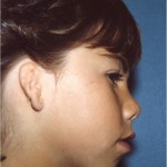
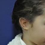
- 6 year old girl with Microtia of her left ear before Rib Graft surgery

-
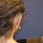
- 1 year post op following Rib Graft surgery on left ear

-

- 21 years post op following Rib Graft surgery on left ear

-
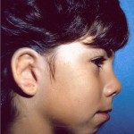
- 8 year old boy with Microtia of the left ear before Rib Graft surgery

-
- 2 years post op following Rib Graft surgery on the left ear
-
- 26 years post of from Rib Graft surgery on the left ear

-
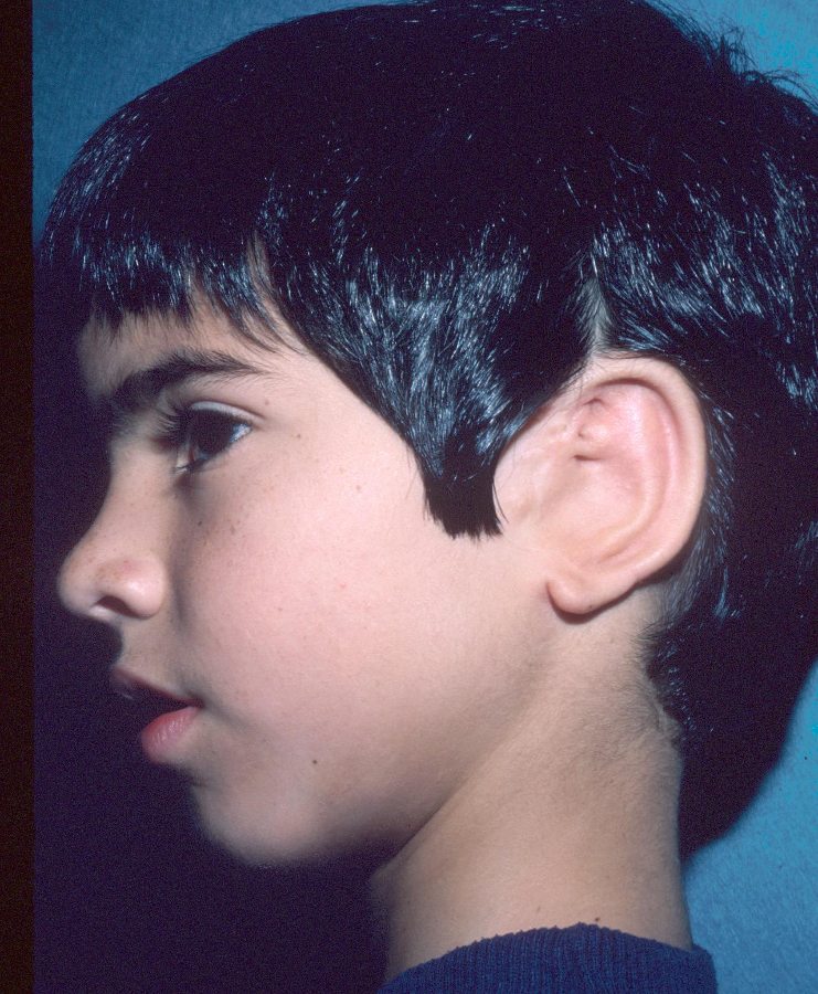
- 9 year old boy with Microtia of the right ear before Rib Graft surgery

-
- 2 years post op following Rib Graft surgery on the right ear

-
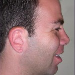
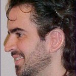
- 30 years post op from Rib Graft surgery on the right ear

The following images are also of one of Dr. Brent’s patients:
-
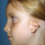
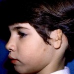
- 8 year old girl with Microtia of her left ear before Rib Graft surgery

-
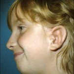
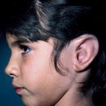
- 1 year post op following Rib Graft surgery on the left ear

-
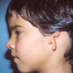
- 4 years post op following Rib Graft surgery on the left ear

-
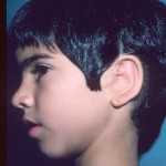
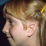
- 12 years post op following Rib Graft surgery of the left ear

-

- 21 years post op following Rib Graft surgery of the left ear

Permission to post the following Rib Graft images has been granted by Dr. Arturo Bonilla.
-

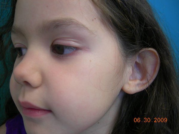
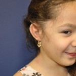
- Girl with right ear Microtia and Atresia (front view)

-
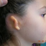
- Girl with right ear Microtia and Atresia (right side view), typical Grade III Microtia

-

- Left side view of non-Microtic left ear
-
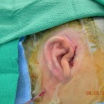
- Immediately following the 1st stage of Rib Graft surgery (right ear)

-
- First post op visit after ear lobe reconstruction with Rib Graft surgery

-
- Elevation stage with Medpor/OmniPore wedge (view 1)

-

- Elevation of ear with Medpor/OmniPore wedge (view 2) following Rib Graft surgery

-
- Post op visit elevation stage (behind the ear view), right ear

-
- Post op visit elevation stage (lateral view), right ear

-
- Front view after tragus reconstruction, right ear

-

- Lateral view after tragus construction (note shadow effect after deepening of conchal bowl), right ear

Below are images from Dr. Bonilla showing how he carves away a small block from the Medpor/OmniPore framework that he inserts behind the ear during the elevation stage.
-
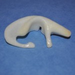
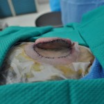
- This is what the Medpor/OmniPore ear framework looks like. Dr. Bonilla will carve a wedge-like piece from the Medpor framework and insert it behind the ear during the elevation stage.

-
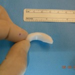
- This is the wedge shaped piece of Medpor/OmniPore material that is carved away and used to be inserted behind the hear during the elevation stage by Dr. Bonilla.

-
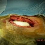
- Medpor/OmniPore wedge being inserted behind ear during the elevation stage with Dr. Bonilla

Interested In Seeing What a Rib Graft Ear Looks Like That is Older Than 10 Years?
Permission to post the following Rib Graft images has been granted by Dr. Arturo Bonilla.
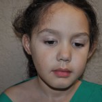
-
- 6 year old girl with left ear Microtia and Atresia before Rib Graft surgery

-
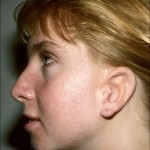
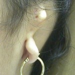
- Now, 14 years post op Rib Graft surgery (Rib Graft surgery was performed at the age of 6)

-
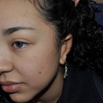
- 7 year old girl with left ear Microtia and Atresia before Rib Graft surgery

-

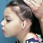
- Now, 14 years post op following Rib Graft surgery, left ear


Leave a Comment
You must be logged in to post a comment.