* This information was gathered by directly speaking with surgeons, families and individuals who have already had surgery, and through personal research. The remaining information has come from noted sources.
What is Microtia?
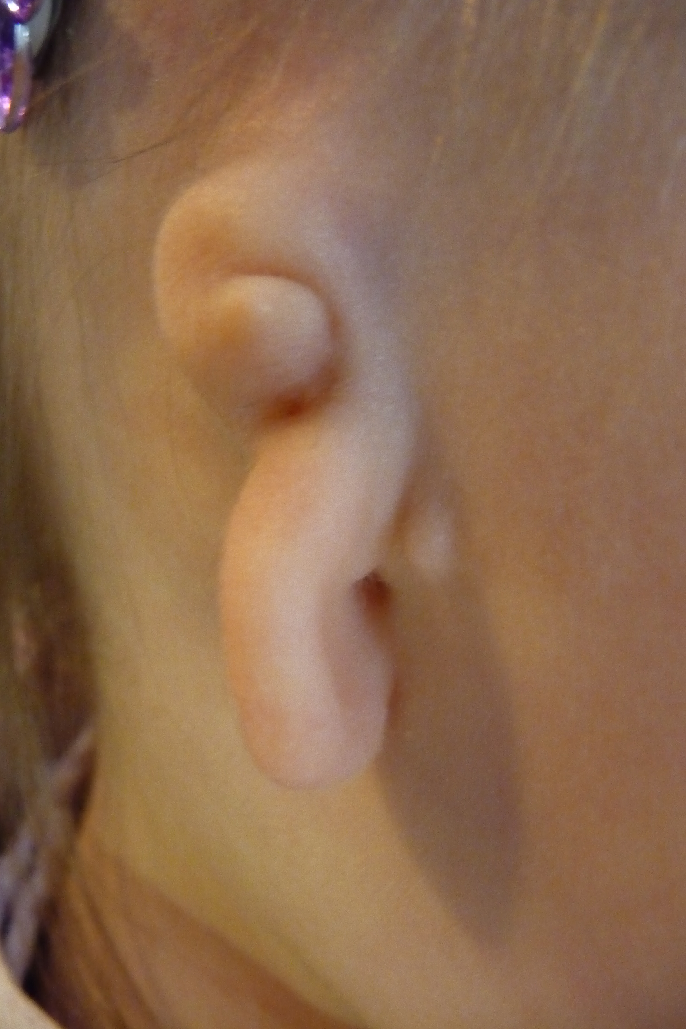
Grade III Microtia and Atresia of the right ear.
Microtia, is a congenital anomaly affecting the outer ear (pinna) where the ear does not fully develop during the first trimester of pregnancy. A Microtia ear is often smaller in size, can have a peanut shaped appearance, only have a small nub or lobe present, or be completely absent at birth. Microtia is Latin for the words micro and otia, meaning “little ear.” Microtia can affect one ear (unilaterally) or both ears (bilaterally). Microtia occurs in every 1 out of 6,000 to 12,000 births. The right ear is more commonly affected. Microtia is often accompanied by Atresia.
Classification:
There are four grades of Microtia:
Grade I: A smaller version of a typical sized ear, still having the same physical characteristics of a typical sized ear, and containing a small but present external ear canal.
Grade II: A partially formed outer ear with very small or narrow ear canals. The ear canals may be very narrow or closed (canal stenosis) producing a conductive hearing loss.
Grade III: Absence of the external ear with a small peanut shaped structure (some cartilage with mostly ear lobe) and an absence of the external ear canal and ear drum (known as aural atresia).
Grade IV: Absence of the complete ear (anotia).
Is Microtia genetic? Yes and No.
No, there are many families who give birth to a child with Microtia when no one on either side of the family has ever had Microtia as far back as history can show. Microtia does not appear to be present in the bloodline for many families. It appears to be a randomly occurring event or anomaly that happens during early development. Families can have a child with unilateral Microtia (right or left ear) or have both ears (bilateral Microtia) affected. One child may be born with Microtia where his/her twin sibling may not show any signs of having Microtia.
Yes, Microtia has shown up in past family generations. Sometimes, an aunt or uncle or a grand parent or cousin may have had Microtia in the past and another family member is born carrying on that same genetic trait. Microtia may be present in only one of two twins or in both of the same set of twins. Microtia may be a trait that a grandfather once had and appears to be passed on to his son and then onto that son’s daughter. Microtia can be passed on by both the maternal and fraternal side. Some relatives who have passed along the Microtia gene (if it is hereditary) may have their right ear affected and then another family member may have their left ear affected. One family member may even have bilateral Microtia when everyone else showed only unilateral signs of Microtia. Although it may appear to be passed on in generations, it can vary.
What if I already have Microtia or already have a child with Microtia, could I have another child with Microtia? It is always possible only because we just don’t know how random Microtia is or how often it can occur in some families who already have it. According to an article written by Dr. Burt Brent, “Incidence of Microtia” there is the possibility of an increased 5% chance of having another child with Microtia if you have already had one. However, even if you do not end up having a child with Microtia, it is possible that one of the children your children has may have a child of their own with Micrtoia or one of your grand children may have a child with Microtia. No one really knows. Live your life how you want to live it. Worst case scenario is that you do have a child with Microtia. Well, you already know what Microtia is, so embrace it. If interested, there are surgical options that can be considered.
There is new evidence showing that Microtia has genetic markers. Although it appears to be in certain ethnic backgrounds (such as Ecuadorian, Hispanic, and Native American cultures), it also appears to be completely random in many others. Some ethnic backgrounds, such as in African American ethnicities, Microtia is very rare. Ecuadorians have a much higher occurrance rate at a 1 in 1,200 chance of having children born with Microtia.
* There are new findings in genetic research in regards to what causes Microtia. You can read about them here and here. Even with new findings on genetic research, there may not be a test available to test for Microtia and Atresia. This is something a genetics counselor or specialist can help determine when you show them the above articles. Otherwise, this is what genetics expert, Dr. Dan Quiat (who worked on this genetics research in collaboration with Ear Community) suggests: “As for a blood test, I would recommend each person discuss with a geneticist what testing might be appropriate. This will differ based on the presence of isolated microtia, CFM, family history, or other conditions that might guide choice of testing. I think we are still in the early days of understanding the genetics, and hopefully in another 5 years there will be a more comprehensive gene ‘testing panel’ to offer all patients.” (October 2022).
How does Microtia affect my child?
Microtia affects the outer ear only. Often times, Microtia is accompanied by Atresia (absence of an ear canal and is often associated with a hearing loss), or Hemifacial Microsomia (where one side of the face has a shorter jaw bone pulling the face slightly upward) or by other associate syndromes or genetic characteristics such as Treacher Collins or Goldenhar Syndrome. However, Microtia does not affect a child’s intelligence, their motor skills or their shot at a normal life to live. As many of us overcome the initial shock we experience after delivering our children, we soon realize how lucky we are as we soon realize that there are other children in our world who are born with life threatening diseases and chronic lifetime ailments. Microtia affects only the ear itself. As our children develop, they will get use to their little ears. Some may not mind their ears as much as others who may not like them much. Try and raise your child with as much confidence and self-esteem as possible for those days where they may feel upset about their ear because of someone questioning it or being teased over their ear. You may just have to instill a little more pride in your child in order to help them get through days like these. Also, if your child struggles with a hearing loss as well, they can still hear with the use of a bone conduction processor. Even if you do not choose to aid your child with an assisted hearing device, help them out a little bit by talking clearly and loudly enough so they can hear you. You may even choose to help your child learn some sign language to help give them that extra opportunity to communicate and not miss out if they can not pick everything up in their environment. Bottom line is that Microtia will only affect your child if you allow it to. Help your child realize that he/she is no different than anyone else and that Microtia can’t get in the way of their life if they don’t allow it to.
How should I raise my child with Microtia?
Raise your newest family addition just as you would raise any other child. You may have to keep an eye on their hearing loss and make sure they are hearing everything they need to be hearing within their environment, but your child will develop into a typical healthy adult. There may be some safety concerns to watch out for regarding traffic if your child does have a hearing loss. Look into any and all options that can help your child get the most out of life, especially options that can help them hear better. Try not to draw attention to their little ear and raise them with confidence as they may experience some bullying. Just be there for them as any parent would be for any one of their children in times like these. A Microtia ear is not “broken”, so it is not something that has to be “fixed.”
Can Microtia be detected in an ultrasound? No.
Typically, sonographers do not focus on looking for Microtia or the details of the ears. Sonographers focus more on the organs of the baby, to make sure everything is developing correctly. Ultrasounds are used to detect abnormal features such as in Down’s Syndrome or mis-shapen limbs, along with general blood screening if requested, etc… Even in a 3D and 4D ultrasounds and Quad Screenings, Microtia is typically not picked up on the sonogram. It is possible that you can ask the sonographer to look for more details as ultrasound technology today has improved greatly. However, this would only notify you that your child would have Microtia. An ultrasound can not prevent Microtia from happening nor can it fix it.
How do I know if my child is deaf or hard of hearing?
Most often, Microtia is accompanied by Atresia (the absence of an ear canal resulting in a loss of hearing). Following the birth of your child or when adopting your child, your child will refer or fail the new born hearing screening. Once your child fails the newborn hearing screening you can take your child to an ENT and an audiologist to have further hearing exams that will reveal how much of a hearing loss your child has. With the help of an ENT and an audiologist, you should be informed about which options can help your child best.
Should we have a CAT scan?
Only if your pediatrician believes your child should have a CT following birth, should you have one. Some times, following the delivery of a child, the child may have other associated syndromes and disorders that the doctor may like to rule out. The doctor may also choose to have a CT taken in order to be certain there is no head trauma or any other associated issues related to brain issues, especially if the child is not responding very well. However, it is strongly suggested by surgeons that a newborn child or a child or younger age not be subjected to a CT until they are older due to the amount of radiation exposure being emitted. The main reason for waiting until the child is older is that CT scans do not always pick up everything. It is also possible that at the time of CT being taken, your child is still developing and therefore, the tiniest bones may not be clearly visible. This would mean your child would now have to be subjected to a second, additional CAT scan. Wait if you can. Many canalplasty (Atresia repair) surgeons suggest waiting to have a CT taken when your child is closer to having ear reconstructive surgery. CT scans are particularly of use in finding out if your child is a candidate for canalplasty. Otherwise, ask your pediatrician or surgeon if a CAT scan is absolutely necessary and if it can wait until later.
How many people have Microtia?
Updated statistics as of 2022:
Approximately 850,000 people live in the world with Microtia and Aural Atresia.
1 in every 6,000 babies are born in the United States with Microtia and Atresia (this may vary with Microtia and Atresia or one or the other).
Approximately 600 babies are born every year in the United states.
There are approximately 58,000 people living in the United States with some form of Microtia and Atresia.
Below are some examples to show you how we came to these numbers:
Studies and findings to date show that Microtia occurs approximately 1 in every 6,000 chances of happening in the United States. You will find online that Microtia happens anywhere from 1 out of 5,000 to 1 out of 12,000. This may be because newer statistics and more recently conducted research hasn’t been updated online for this rare cause. Also, some statistics may be blended including other countries and regions where these numbers to differ.
Let’s say that Microtia has a chance of occurring 1 in 9,000 times.
As of November 2018, there were approximately 7.6 billion people living in the world.
United States statistics…
* Of those 7.6 billion people, approximately 327 million are living in the United States. With Microtia having a 1 in 9,000 chance of occurring, that means that there are approximately 36,000 people living in the United States with Microtia.
United Kingdom statistics…
* Again, of those same 7.6 billion people living in the world, approximately 66 million are living in the United Kingdom. With Microtia having a 1 in 9,000 chance of occurring, that means that there are approximately a little more than 7,000 people living in the United Kingdom with Microtia.
Australian statistics…
* Again, of those same 7.6 billion people living in the world, approximately 24 million are living in Australia. With Microtia having a 1 in 9,000 chance of occurring, that means that there are approximately 2,600 people are living in Australia with Microtia.
So, out of 7.6 billion people living in our world today, approximately 844,000 were born with some degree of Microtia.
What specialists should I be taking my child to?
Neonatologist: Your child may be seen by a neonatologist in conjunction with your pediatrician and obstetrician to help evaluate your child for a proper diagnosis and to help answer questions you may have following the birth of your new baby.
Otolaryngologist and Audiologist: Your child should first be seen by an Otolaryngologist (ENT/ear-nose-throat specialist) and an audiologist for hearing checks and to find out at what level your child’s hearing loss is. You should automatically be referred to see both of these specialists following the newborn hearing screening. Especially, if your child “refers on the newborn hearing screening” which means they did not pass the hearing test. Ask your ENT and audiologist about what options are available that can help your child hear better. Set up quarterly visits to stay on top of hearing and ear infections at least through age 3. Then, consider a hearing test twice annually until age 5 with an annual hearing test after age 5.
Craniofacial Surgeon and Oral Surgeon: If your child has Craniofacial Microsomia (also known as Hemifacial Microsomia), you may want to consider setting up an appointment with a craniofacial surgeon and an pediatric oral surgeon/oral surgeon. Both of these specialists should be part of a craniofacial team that should be suggested you meet with following your child’s birth. Begin inquiring about information that can help your child as he/she develops and ask about the possibility of surgery and at what age your child should begin surgery if surgery is being considered. Such surgeries may include craniofacial repair to lengthen the jaw line, evening out facial asymmetry, tooth extractions and braces. A pediatric dentist or pediatric oral surgeon is a good friend to have during childhood for children with Microtia. A plastic surgeon on the craniofacial team can also help with additional options for Microtia including facial inserts that can help even out the symmetry of the face and also the option for Botox injections.
Pediatrician or General Family Physician: These are the doctors that your child will see most of their life. Make sure they are kept in the loop on information from other medical specialists by having copies of evaluations and checkups, xrays, etc…
Microtia Reconstructive Surgeon and a Canalplasty Surgeon: If you are interested in finding out more about surgical options for your child, you may want to consider scheduling an appointment with an expert reconstruction specialist or plastic surgeon who specializes in facial reconstruction surgery and Atresia repair. Ask questions so you can learn what options are available for your child and how to begin planning for surgery including appropriate ages, schedules that work best for you when planning for the future, best options and candidacy as well as insurance and any upfront or out of pocket co-pays. Many families end up fund raising if a surgeon’s office does not accept insurance and is private pay.
Anaplastologist: A prosthetic ear specialist who can help you review the option for a prosthetic ear as an option for Microtia.
Additional specialists you may choose to see for Microtia and Atresia are:
Counselor/Social Worker/Therapist – many children and families struggle to understand why this happened to them. Just simply having someone to speak with can help greatly.
Speech Therapist – due to Aural Atresia causing hearing loss when accompanying Microtia, speech delays may result.
Physical Therapist – while Microtia doesn’t typically cause the need for physical therapy, some children may be born with low muscle tone and be delayed in walking. Also, some children may develop torticollis as well where a physical therapist can be of help.
Occupational Therapist – while Microtia doesn’t typically cause the need for occupational therapy, many children may need a little extra help during the infant/toddler years where an occupational therapist can be of help.
Behavioral Psychologist/Developmental Psychologist – may be able to help if your child with Microtia is experiencing challenges with being on par with their peers, be of a smaller stature in size, behind in milestones in general. This may be due to other associated syndromes or challenges your child may also have been born with in addition to Microtia.
Geneticist: You may choose to consult with a geneticist regarding finding out any additional information that may help to explain how or why Microtia, Atresia, Craniofacial Microsomia/Hemifacial Microsomia occurred. Currently, there is not a specific genetics test that can be conducted for Microtia and Atresia. However, a geneticist counselor can help you discover if your child’s Microtia and Atresia are related to other syndromes. Some syndromes that Microtia and Atresia are often associated with or accompanied by are: Goldenhar Syndrome, CHARGE Syndrome, Treacher Collin’s Syndrome or BOR Syndrome. Craniofacial Microsomia and Hemifacial Microsomia are often associated with those who have Microtia.
* There are new findings in genetic research in regards to causes of Microtia. You can read about them here and here. Even with new findings on genetic research, there may not be a test available to test for Microtia and Atresia. This is something a genetics counselor or specialist can help determine when you show them the above articles. Otherwise, this is what genetics expert, Dr. Dan Quiat (who worked on this genetics research in collaboration with Ear Community) suggests: “As for a blood test, I would recommend each person discuss with a geneticist what testing might be appropriate. This will differ based on the presence of isolated microtia, CFM, family history, or other conditions that might guide choice of testing. I think we are still in the early days of understanding the genetics, and hopefully in another 5 years there will be a more comprehensive gene ‘testing panel’ to offer all patients.” (October 2022).
Can someone with Microtia have reconstructive ear surgery?
Yes, there are two surgical options for ear reconstructive surgery for Microtia, Rib Graft or auricular ear reconstruction and porous polyethylene, with a third reconstructive option coming in the future.
Surgical Options for Microtia Repair (reconstructive surgery):
1. Rib Graft Surgical Technique:
Known as the “Gold Standard” for ear reconstructive surgery and has been around since the 1920’s. A three to four stage surgery that involves using a section or a piece of rib cartilage (rib graft) and a section of your skin needed to cover the ear (skin graft). Together the rib cartilage and skin graft are used to reconstruct an outer ear. The 1st stage involves the harvesting of the rib cartilage (requires a 1” to a 1.5” length incision) and then carefully sculpting the rib cartilage into a newly shaped framework that is shaped like an ear. The newly shaped framework is then sewn into a skin flap or skin pocket beneath the scalp on the skull (where the ear will be located). The 2nd stage involves creating the earlobe. The 3rd stage involves making a slit behind the ear to careful release the ear from beneath the scalp skin, lifting it away giving it projection or a high profile look. A skin graft is then used to help covering up the rest of the ear (back side of the ear). This is known as the elevation stage. The third stage is where the tragus (little cartilage like flap located in front of the ear canal) is constructed and any other symmetrical issues with the ear are then corrected at best. This stage of the surgery also helps create a pseudo ear canal with a shadowing affect that provides a depth perception look as if an ear canal exists (if a patient is not a candidate for canalpasty (atresia repair). Any additional stages are typically only required for tweaking of the ear itself, such as tweaking the shape of the ear more or the earlobe. Since a rib graft ear is made of living tissue from the body, the ear will grow with the individual and will be undersized when compared to the existing non-Microtic biological ear so that it can grow to approximately the same size of the existing ear. The newly reconstructed rib graft ear will be slightly stiffer than the biological ear as the rib cartilage tissue used is thicker or stronger than ear cartilage, although the ear appears to physically be similar in thickness to the human eye. The ear will feel pain, experience bleeding, and heal. The earliest age to consider Rib Graft surgery is age five. More common ages for Rib Graft surgery range between six and ten years of age.
Note: Some surgeons can achieve ear reconstructive surgery in fewer than three or four stages. Surgeon skill and technology has advanced over the years. Some surgeons can now perform a rib graft procedure in as little as 1 to 2 states of surgery. Surgery time for each stage ranges anywhere from one hour to five hours or more, depending on the stage and what needs to be done to the ear for best results. Longer surgery times and additional stages may be required for children having Goldenhar Syndrome and Treacher Collins because of the challenge of centrally positioning the ear and working with a lower or higher hair line. Some history on surgical techniques began with the “Nagata” technique, named after Dr. Satoru Nagata of Japan, performed Rib Graft surgery in only two stages as well as the “Firmin” technique, named after Dr. Francoise Firmin of France. Surgeries typically performed in three stages or so most-likely utilize the “Brent” technique, named after Dr. Burt Brent of California. However, many surgeons today have developed their own surgical techniques anywhere from combining a mixed skill, their own or utilizing equipment to help carve an ear such as Auryzon by Reconstrata that cuts a consistent ear framework, saving hours of time in the OR when vs using the Gold Standard of stitching an ear framework from multiple sections of rib.
2. Medpor/Omnipore/SuPor Surgical Technique:
A surgical technique that eliminates the additional stages required for harvesting rib cartilage for rib graft procedures. Medpor/Omnipore/Supor surgery utilizes a synthetic pre-made framework known as a porous polyethylene ear frame work. This pre-formed ear framework comes already formed for the surgeon that can be manipulated to match the shape of the non-Microtic ear (for example, adding a dimple). The Medpor, Omnipore or Supor surgical techniques utilize a porous polyethylene material, the same material that has been used in facial craniomaxillofacial surgeries for over fifty years. This surgery can be achieved in just one stage. Surgery time can range anywhere from eight to twelve hours depending on the complexity of the surgery. A second stage may be required to tweak the earlobe if needed. The ear is constructed to be slightly larger in size (around .5mm or less in size) than the already existing non-Microtic ear, allowing the biological ear to grow to the same approximated size of the newly reconstructed Medpor ear. A Medpor ear will be stiffer than a rib graft ear as the material the synthetic framework is made up of is a bit thicker or stronger than ear cartilage, although the ear appears to physically be similar in thickness to the human eye. A Medpor, Omnipore, Supor ear does become fully vascularized where blood vessels become integrated throughout the porous holes within the polypropylene frame. This allows the ear to feel pain, experience bleeding, and heal. The earliest age to consider Medpor surgery is three to four years of age.
3. Regenerated Ears and 3D printing:
Regenerated ears are coming! Some clinical trials have already begun as of 2021. Some regenerated ear options include 3D printing. Some articles where you can read about regenerated ears can be found here and here.
Are there other options for Microtia other than surgical options? Yes.
1. The Do Nothing Option:
You can always choose to accept your Microtia ear(s) and choose to do nothing. Keep your little ear just the way it is, love your ear and embrace your ear. Many individuals like their ears just the way they are. They believe their ear is a part of them and they and their family do not wish to change their ear in any way, shape or form. In fact, some individuals with Microtia adore their little ears and show them off with piercings and wearing their hair short. Some even believe their little ears have added to their character. You do not have to do anything to your ear. If you have adjusted to life with it and don’t mind your ear, then it is absolutely fine to do nothing. You should never let anyone push you into making a decision that do not agree with or do not want to make yourself. Little ears are beautiful and so are the individuals wearing them.
2. Prosthetics:
Choosing to wear a prosthetic ear can be an excellent option to consider instead of having surgery performed. Prosthetic ears today look amazing and realiztically real. They can easily be worn by either adhesive (glue) or by being anchored on with magnets or a clip bar. In fact, around 2015, depending on the size of the Microtic ear, the ear remnant no longer has to be removed in order to wear a glue on prosthesis. A prosthetic ear can be fit to cover the Microtic ear if you are not yet comfortable with removing your existing ear. You can swim with a prosthetic ear and you can sleep with a prosthetic ear (although this may degrade the quality of the ear over time). As long as you follow the suggested daily cleaning instructions, wearing a prosthetic ear can be quite easy. Thanks to advancements in technology, today, many prosthetic ears are often 3D printed to perfection, even though anaplastologists are amazing artists to begin with.
Can Microtia affect my child’s growth? No.
Microtia usually does not affect the growth of a child. Some children are just smaller in stature than others. Some of the children who grow up smaller in size are often the ones who experience growth spurts in middle school and high school and then are taller than the other children. Just because a child has a smaller stature does not mean that Microtia is to blame or that Microtia has stunted the growth of your child. We often worry too much and look for things to point the finger at. It is possible that your child may have another undetected associated syndrome with their Microtia that may be causing their petite stature. Again, this would not be because of Microtia, but because of the newly associated syndrome. If you are concerned about your child’s growth, consider contacting a specialist in endocrinology. Please keep in mind that every child is different and they are often different from their siblings too. It is very hard to tell sometimes. Often times, your child’s pediatrician will say your child is fine as long as they are healthy and growing in proportion. Ask your pediatrician questions if you are concerned.
There have been some suggestions for doing sleep studies, especially if your child has Craniofacial Microsomia/Hemifacial Microsomia. It is possible that sleep apnea can affect growth. Ask your ENT about having a sleep study scheduled for your child. New research and findings are always just around the corner.
What are some theories explaining how Microtia can happen?
1. A slight lack of oxygen or drop in oxygen levels during pregnancy within the first trimester may caused the ear to cease in development. During the 1st trimester, the heart, kidneys, and ears all develop at the same time. These organs can be affected during the same time. This is the reason is it often suggested to have renal ultrasounds and EKGs on our children following delivery.
2. It is possible that the the umbilical cord some how slightly rubbed up against the fetus during development during the 1st trimester. Although the fetus is only about two inches or less in length and about the size of a lime at this age, it is still possible that this theory can happen. An example would be when children are born with under-developed limbs (not related to drugs) because the umbilical cord some how was touching or wrapped around the limb and the limb ceased in development.
3. Taking drugs and alcohol during pregnancy can cause Microtia and Atresia. Though, this may be true, chances are that the drugs and alcohol also caused other medical issues too and Microtia or Atresia is just one of the affects from the drugs and alcohol. Fetal Alcohol Syndrome has been know to cause Microtia in some individuals. Microtia can also result from taking Accutane (isotretinoin) and Methamphetamines during pregnancy.
4. See new findings about genetics in Microtia under our RESOURCES menu tab here.
What are some myths about Microtia?
1. Microtia is a disease. No, Microtia is not a disease, nor is it a virus or a contagion. You can not catch Microtia from anyone or from visiting a foreign country. It is a congenital deformity that just happens.
2. Microtia is caused by taking fertility drugs, such as Clomid (Clomiphene). There is no evidence proving that the use of fertility drugs can cause Microtia.
3. Microtia is caused by drinking certain types of milk. There is no evidence proving that by drinking milk in general (any and all types) can cause Microtia. There is also no proof that by eating or drinking anything from your environment can lead to causing Microtia.
* Even though some research has been tied to Microtia with diabetes or from taking Accutane, just as many people who have had babies with Microtia do not have diabetes or took Accutane. Also, some believe that bad viruses such as RSV cause Microtia when so many other mothers didn’t have this or were even sick during their pregnancies. These are some related examples that have come across in research.


Leave a Comment
You must be logged in to post a comment.